Essential Otolaryngology: Head and Neck Surgery
8,890.00₹ 3,995.00₹
For more than four decades, K.J. Lee’s Essential Otolaryngology has been the premier international guide to otolaryngology. This classic reference delivers top-to-bottom coverage that spans the entire discipline and provides an easy, at-a-glance review of the field’s must-know information. The tenth edition features quick-access bulleted text, 150 illustrations, 100 tables, and thoroughly updated chapters that bring you up to speed with today’s practice of otolaryngology.
Features:
- The latest clinical information to help you treat dozens of conditions involving the head and neck, including:Balance disorders,Facial nerve paralysis,Congenital hearing loss,Cleft lip and palate,Infections of the temporal bone,Balance disorders,Facial nerve paralysis,Tumors of the larynx,Sinusitis,Cysts and tumors of the jaw
- Coverage of nutrition, fluid, and electrolytes; facial plastic surgery; anesthesia for head and neck surgery; and pharmacology and therapeutics
- The perfect otolaryngology primer and the ideal board review resource – in one compact volume
- An increased number of diagnostic algorithms to enhance clinical decision making
Katzung & Trevor’s Pharmacology Examination and Board Review
3,990.00₹ 2,295.00₹
From the authors of pharmacology’s #1 textbook, Katzung & Trevor’s Basic & Clinical Pharmacology, Twelfth Edition, this review delivers a clear, concise review of fundamental concepts backed by more than 1,000 review questions and answers. The chapter-based approach facilitates use with course notes or larger texts.
Outstanding learning aids include:
- Short discussion of the major concepts that underlie basic principles or specific drug groups
- Explanatory figures and tables
- Review questions followed by answers and explanations
- Drug trees in drug-oriented chapters that visually organize drug groups
- A list of high-yield terms and definitions you need to know
- Skill Keeper Questions that prompt you to review previous material to understand links between related topics
- A checklist of tasks you should be able to perform upon completion of a chapter
- Summary tables that list the important drugs and include key information about their mechanisms of action, effects, clinical uses, pharmacokinetics, drug interactions, and toxicities
- Two comprehensive 100-question examinations followed by the correct answers and rationales
- Valuable test-taking strategies for improving your test performance
Positron Emission Tomography-Computed Tomography
29,330.00₹ 15,190.00₹
Positron Emission Tomography Computed Tomography: A Disease-Oriented Approach offers Radiologists and Nuclear Medicine specialists a thorough understanding of the clinical application of PET-CT―a groundbreaking modality that provides a powerful fusion of imaging anatomy and metabolic function. Written with a disease-oriented approach, PET-CT examines understanding, using, and interpreting PET-CT imaging in clinical practice. Co-authored by experts in both PET and CT imaging, this text serves as an integrated review of the practical aspects of this new imaging modality while providing comprehensive and evidence-based coverage. This volume covers all clinical entities for which PET-CT can be utilized in today’s modern practice.
Using an integrated disease-oriented approach, PET-CT reviews:
- the diagnostic settings in which PET-CT will prove most valuable
- literature-based evidence for utility, applications, and limitations to each disease
- integrated discussion of the CT findings that will bear on the PET interpretation and vice versa
- “next steps” in the clinical evaluation of a patient (i.e., additional imaging studies indicated)
Positron Emission Tomography Computed Tomography also includes a CD packed with every image from the book. With over 340 high resolution photos this makes a perfect addition to both in-depth study and PowerPoint slide presentations.
Body CT The Essentials
5,810.00₹ 3,990.00₹
Body CT: The Essentials delivers an up-to-date, detailed, and practical review of CT imaging of the chest, abdomen, and pelvis. It will prove especially valuable to trainees in diagnostic radiology and practicing radiologists with an interest in body imaging.
Primarily organized by organ system, Body CT: The Essentials also includes important technical chapters that review intravenous contrast administration, scan parameters, and radiation physics that enable you to perform quality studies with minimum patient radiation exposure.
Each organ-specific chapter incorporates the latest advances in CT imaging and recommendations or guidelines for imaging, as well as follow-up findings. Tables found within the chapters include differential diagnosis, and each chapter concludes with suggested readings for a more detailed discussion of the topic.
Here’s why this is the perfect CT primer:
- Enhanced by more than 450 images
- Emphasizes the appropriateness and role of CT relative to other imaging modalities and protocols
- Includes coverage of the latest technologies such as cardiac CT, CT colonography, and CT enterography
- Focuses on the most practical concepts related to generating a concise, accurate differential diagnosis and relevant report
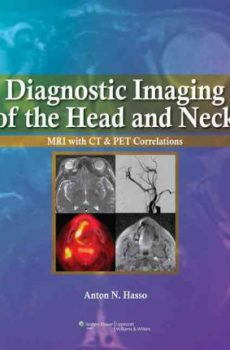
Diagnostic Imaging of the Head and Neck MRI with CT & PET Correlations
15,400.00₹ 3,490.00₹
A single-authored, clinically oriented text on imaging of the head-and-neck, frequently a difficult area for radiology residents and general radiologists to master. Readers will find key diseases highlighted and a guide to differential diagnosis of various conditions. Though the primary image focus is on MRI, correlations with CT and PET images and strong coverage of anatomic variants–to distinguish those from the presence of disease–are major strengths of the book. Other features include excellent image quality, diagrams and tables. While this text does not replace the need for a comprehensive text, it should be an essential resource at the reading station and on rotation.
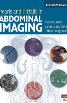
Pearls and pitfalls in abdominal imaging
10,400.00₹ 4,795.00₹
Research consistently suggests that 1.0 to 2.6% of radiology reports contain serious errors, many of which are avoidable, and it is clear that all radiologists can struggle with the basic questions as to whether a study is normal or abnormal. Pearls and Pitfalls in Abdominal Imaging presents over 100 conditions in the abdomen and pelvis which can commonly cause confusion and mismanagement in daily radiological practice, providing a focused textbook that can be readily used to avoid wrong diagnoses and prevent incorrect management or even malpractice litigation. It includes 700 figures and covers all the major modalities including CT, PET/CT and MRI. The pearls and pitfalls include artifacts, anatomic variants, mimics, and a miscellany of diagnoses that are under-recognized or only recently described. Conditions are presented in anatomic order from diaphragm to the symphysis pubis, with grouping by location and organ system.
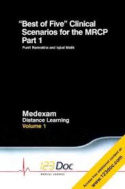
Best of Five Clinical Scenarios for the MRCP
6,990.00₹ 4,490.00₹
The recent change in examination format has made the revision books and materials published over the years largely obsolete. This new book is based on the new form of MCQ – ‘Best Of Five’ Clinical Scenarios, matching the style and standards set by the College, and is designed to provide readers with a complete distance-learning package. This first volume in the series can be accessed online to experience all the benefits of the 123Doc’s celebrated e-courses. It covers eight of the most important specialities seen in the MRCP exam: Cardiology, Dermatology, Gastroenterology, Haematology, Infectious Diseases, Nephrology, Neurology and Respiratory Medicine (Volume 2 will cover the remaining specialties). Within each chapter, ‘hot-topics’ from the speciality are illustrated by 2 questions, with detailed teaching notes and lists of ‘key points’ to help you further master the subject.Published in association with 123Doc Medical Courses.
Essentials of Pediatric Radiology
39,990.00₹ 13,790.00₹
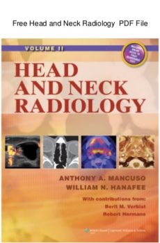
Written and edited by acknowledged masters in the field, this two-volume full-color text is the most comprehensive and current reference on head and neck radiology. It features more than 10,000 images and covers every disorder in every region of the head and neck.
The first two sections cover applied imaging fundamentals and general pathology, pathophysiology, patterns of disease, and natural history of head and neck disorders correlated with imaging appearance. Subsequent sections focus on specific anatomic regions: the eye, orbit, visual pathways, and cranial nerves III, IV, and VI; sinonasal and craniofacial region including cranial nerve V; temporal bone, posterior skull base, posterior fossa, and cranial nerves VII-XII; infrahyoid neck and cervico-thoracic junction (thoracic inlet); thyroid and parathyroid glands; major salivary glands; nasopharynx; oropharynx; oral cavity and floor of the mouth; larynx, hypopharynx, and cervical esophagus; trachea; hypopharynx; and cervical esophagus. The text covers all current imaging modalities, including plain film, MRI, CT, ultrasound, and nuclear medicine including PET.

Molecular Anatomic Imaging
20,000.00₹ 9,700.00₹
This fully updated Second Edition is perfect for clinicians, as it focuses sharply on clinical PET-CT and SPECT-CT examinations, omitting lengthy physics discussions. The book is now strictly disease oriented and integrates PET-CT and SPECT-CT applications completely. When both techniques are relevant for a disease, they are discussed together; when only one is relevant, it is discussed alone. More than 1,200 illustrations document the use of integrated imaging and provide very useful reference material for interpreting integrated imaging studies.
A bound-in DVD contains over 80 cases to be viewed in three orthogonal planes and different CT windows organized as reference and self-assessment files. The cases provide excellent training and self-assessment material. Readers can use the DVD to enhance the book’s illustrations or to test their abilities in making diagnoses on their own.
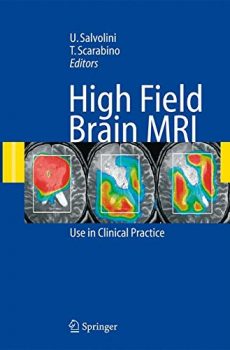
High Field Brain MRI
11,200.00₹ 6,300.00₹
This book describes the development of systems of magnetic resonance imaging using the higher magnetic field strength of 3 tesla, in comparison to the current gold standard of 1.5 tesla. These new systems of MRI make it possible to perform with high spatial, temporal and contrast resolution not only morphological examinations but also functional studies on spectroscopy, diffusion, perfusion, and cortical activation, thus helping research and providing an important tool for routine diagnostic activity. At the same time the new systems offer unparalleled sensitivity and specificity in the numerous conditions of neuroradiological interest.
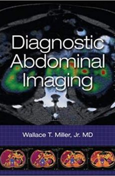
Diagnostic Abdominal Imaging
16,290.00₹ 9,790.00₹
Diagnostic Abdominal Imaging provides a comprehensive review of abdominal diseases based on pattern recognition. Utilizing more than 2,300 images, the book includes discussions of the x-ray, sonographic, CT, MRI, and nuclear radiology features of abdominal diseases. Since accurate imaging diagnosis of diseases can only be achieved with the appropriate clinical history, the characteristic clinical presentations of abdominal diseases are discussed in conjunction with the image findings.
Presented in fifteen organ-based chapters that highlight differentiation of disease on the basis of imaging patterns, Diagnostic Abdominal Imaging discusses the full spectrum of malignant and nonmalignant abdominal disorders. Each discussion begins with the most salient histologic, pathologic, and clinical features of the disorder under discussion. This is followed by a systematic review of the imaging features of the disease as seen by all modalities.
Unlike most radiology texts which are organized by pathology, Diagnostic Abdominal Imaging is organized by imaging appearance―mimicking real-world practice. The book guides you through the process of imaging-based diagnosis and stresses the epidemiological, clinical, and imaging features that allow the most accurate prediction of disease.
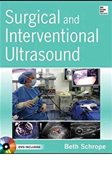
Surgical and Interventional Ultrasound
12,900.00₹ 7,700.00₹
All the guidance you need to enhance your understanding and clinical application of ultrasound
Includes DVD with video of key techniques
Surgical and Interventional Ultrasound offers a thorough survey of image-guided treatments in the OR, in the endoscopy suite, and at the bedside. This one-stop clinical companion spans virtually every kind of surgical and interventional specialty that utilizes ultrasound and delivers high-yield perspectives on using these techniques to ensure accurate clinical decision making.
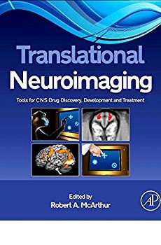
Translational Neuroimaging
13,300.00₹ 7,700.00₹
ranslational Neuroimaging: Tools for CNS Drug Discovery, Development and Treatment combines the experience of academic, clinical and industrial neuroimagers in a unique collaborative approach to provide an integrated perspective of the use of small animal and human brain imaging in developing and validating translational models and biomarkers for the study and treatment of neuropsychiatric disorders. Translational Neuroimaging: Tools for CNS Drug Discovery, Development and Treatment examines the translational role of neuroimaging in model development from preclinical animal models, to human experimental medicine, and finally to clinical studies. The focus of this book is to identify and provide common endpoints between species that can serve to inform both the clinic and the bench with the information needed to accelerate clinically-effective CNS drug discovery. This book covers methodical issues in human and animal neuroimaging translational research as well as detailed applied examples of the use of neuroimaging in neuropsychiatric disorders and the development of drugs for their treatment. Translational Neuroimaging: Tools for CNS Drug Discovery,
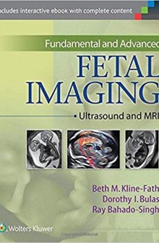
Fundamental and Advanced Fetal Imaging
20,900.00₹ 6,400.00₹
Effectively evaluate obstetric patients with Fundamental and Advanced Fetal Imaging: Ultrasound and MRI! Written by an impressive roster of leading fetal radiologists and maternal-fetal medicine specialists, with additional input from cardiologists, geneticists, and Doppler specialists, this state-of-the-art reference explores how to obtain the maximum information from fetal ultrasound and magnetic resonance imaging, so you can rule out pathologies with confidence – or identify them early enough to initiate the most appropriate interventions. Key Features • Addresses normal anatomy and imaging techniques; screening for normal and abnormal conditions; fetal malformations, emphasizing the essential criteria needed to diagnose each disorder • Explores the major fetal anomalies by organ system, as well as common genetic and chromosomal disorders, outlining their incidence, pathogenesis and etiology, diagnosis with US and MRI, prognosis, and management • Offers abundant imaging examples representing the full range of US and MRI findings, as well as color diagrams and tables to expedite reference Now with the print edition, enjoy the bundled interactive eBook edition, offering tablet, smartphone, or online access to: Complete content with enhanced navigation • A powerful search that pulls results from content in the book, your notes, and even the web • Cross-linked pages, references, and more for easy navigation • A highlighting tool for easier reference of key content throughout the text • The ability to take and share notes with friends and colleagues • Quick reference tabbing to save your favorite content for future use
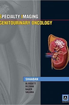
Specialty Imaging: Genitourinary Oncology
9,730.00₹ 4,790.00₹
The first Amirsys publication devoted solely to genitourinary cancers has finally arrived. In Specialty Imaging: Genitourinary Oncology, Dr. Akram Shaaban and team skillfully guide clinicians through the imaging appearances, staging, and treatment techniques for cancers of the kidney, bladder, adrenal glands, urethra, testicle, and prostate. The 130 pages of fast-reading, bulleted text are enriched with well over 450 vivid images, each of which identifies and explains clinically significant factors. The recently published Seventh Edition AJCC cancer staging tables are included to aid the staging discussions, providing definitions for TNM and AJCC prognostic groups for all genitourinary cancers. This is a must-have reference for radiologists, oncologists, and others whose work quality depends on an accurate understanding of genitourinary system cancers.
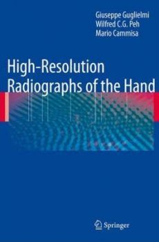
HIGH RESOLUTION RADIOGRAPHICS OF THE HAND
9,600.00₹ 3,790.00₹
Plain radiography is still alive. In many institutions, including ours, conventional radiography has been replaced by digital systems including imaging-plate-based computed radiography and fat-panel detector-based digital radiography. Even for the education of radiation technologists, conventional flm-screen radiography has been de– phasized, and their education is concentrated on digital systems. Spatial resolution of a conventional system is still far better than the current digital systems, although the dynamic range is wider in the latter system. Industrial flm radiography with small grain size and direct exposure has an even higher resolution, and such hi- resolution systems are something we lost in the transition from the conventional system to the current PACS-friendly system. I am pleased to know that Giuseppe Guglielmi and Wilfred Peh have published this textbook of high-resolution hand radiographs that cannot be obtained with any other techniques. Radiography has always been the most important modality in the evaluation of the hand, and, moreover, high-resolution industrial flms are extremely efective in the evaluation of the hand, particularly for assessing subtle erosions. Hands are not just one of the peripheries of the human body. Tey refect conditions of the whole human body. Not only the metabolic status, but also many congenital disorders are manifested in the hand. Radiographic fndings of the hand are ofen specifc, and contribute to the diagnoses a great deal. Tere have been several publications concerning the radiology of the hand, and they have been well accepted.
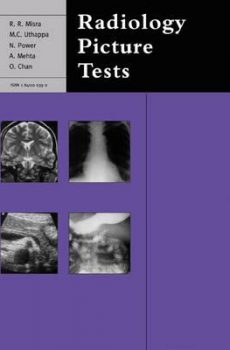
Radiology Picture Tests
3,900.00₹ 2,700.00₹
A comprehensive, systemically organised ‘film viewing’ book designed specifically for FRCR candidates but equally useful to all trainee and junior radiologists as an ‘aide memoire’ and to medical undergraduates and MRCP candidates as a useful means of self-testing their level of knowledge. It is fully illustrated with high-quality, clearly labeled black and white illustrations throughout and the text closely mirrors the way that questions are asked in the FRCR film viewing examination, so as to provide as real an examination experience as possible. Each section of the book contains 20 representative questions, and model answers, providing 200 cases altogether.
Radiologic Differential Diagnosis
4,620.00₹ 2,990.00₹
This is the perfect quick-reference for the busy radiologist and resident. The most up-to-date resource in the field of radiography, this bulleted handbook provides differentiating features for hundreds of diseases, conditions, and injuries. Readers will find 300 illustrations and radiographs, a streamlined find-it-now format, and exceptional value for the price.
The Physics of Clinical MR Taught Through Images
5,300.00₹ 3,300.00₹
Portions of the book have been presented at both the RSNA (Radiological Society of North America, 2003) meeting and the ARRS (American Roentgen Ray Society, 2004) meeting. Awards were given to one of the two exhibits shown at each meeting, with the award at the ARRS being very prestigious (a ‘silver medal’). At the 2004 ARRS meeting there were 229 exhibits presented. One gold medal, three silver medals, six bronze medals and 30 certificates of merit were awarded. The exhibits were also chosen for submission to Radiographics (which was declined, as there were copyright issues). Other sections of the book have been submitted for presentation at the 2004 RSNA meeting, and if accepted there, will also be submitted to the 2005 ARRS meeting.