Working Hours - Mon - Sat 9.00 am to 7.30 pm - 0495 - 2740321, +91 9747580707

NEWS AND EVENTS
  • Here’s why Dan Brown hasn’t written about Hinduism yet in his Robert Langdon novels

    Dan Brown is once again taking on the big questions.…

  • STOP PRESS!

    Kazuo Ishiguro wins the Nobel Prize For Literature The British…

read all News

Radiology

Showing 21–40 of 51 results

  • Handbook of Breast MRI

    7,070.00 4,790.00

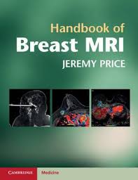
    Breast MRI is no longer the domain of specialised centres; it is now a mainstream diagnostic technique, and an understanding of its applications is essential for any clinician involved with breast imaging. The Handbook of Breast MRI provides core knowledge and clinical guidelines for performing breast MRI in everyday practice. Introductory chapters on breast MRI basics, anatomy and pathology are followed by detailed chapters on the use of MRI in screening, staging, problem-solving and MRI-guided interventions, each containing diagnostic algorithms, tables and lists for quick access to key diagnostic information. Each chapter also contains a selection of self testing questions, and numerous Appendices concisely summarise tumour classification and current breast cancer treatment options. The Handbook of Breast MRI is an invaluable practical diagnostic resource for radiologists, surgeons, oncologists and all clinicians involved in breast cancer management.

  • High Field Brain MRI

    11,200.00 6,300.00

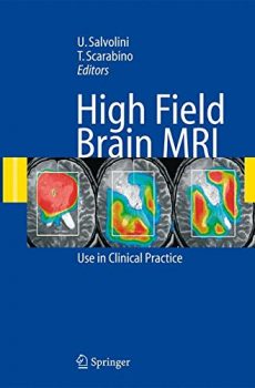
    This book describes the development of systems of magnetic resonance imaging using the higher magnetic field strength of 3 tesla, in comparison to the current gold standard of 1.5 tesla. These new systems of MRI make it possible to perform with high spatial, temporal and contrast resolution not only morphological examinations but also functional studies on spectroscopy, diffusion, perfusion, and cortical activation, thus helping research and providing an important tool for routine diagnostic activity. At the same time the new systems offer unparalleled sensitivity and specificity in the numerous conditions of neuroradiological interest.

  • HIGH RESOLUTION RADIOGRAPHICS OF THE HAND

    9,600.00 3,790.00

    Plain radiography is still alive. In many institutions, including ours, conventional radiography has been replaced by digital systems including imaging-plate-based computed radiography and fat-panel detector-based digital radiography. Even for the education of radiation technologists, conventional flm-screen radiography has been de– phasized, and their education is concentrated on digital systems. Spatial resolution of a conventional system is still far better than the current digital systems, although the dynamic range is wider in the latter system. Industrial flm radiography with small grain size and direct exposure has an even higher resolution, and such hi- resolution systems are something we lost in the transition from the conventional system to the current PACS-friendly system. I am pleased to know that Giuseppe Guglielmi and Wilfred Peh have published this textbook of high-resolution hand radiographs that cannot be obtained with any other techniques. Radiography has always been the most important modality in the evaluation of the hand, and, moreover, high-resolution industrial flms are extremely efective in the evaluation of the hand, particularly for assessing subtle erosions. Hands are not just one of the peripheries of the human body. Tey refect conditions of the whole human body. Not only the metabolic status, but also many congenital disorders are manifested in the hand. Radiographic fndings of the hand are ofen specifc, and contribute to the diagnoses a great deal. Tere have been several publications concerning the radiology of the hand, and they have been well accepted.

  • Image Guided Core Biopsy of the Breast: A Practical Approach

    4,500.00 1,990.00

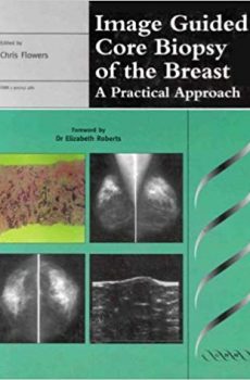
    Image Guided Core Biopsy of the Breast is the first book to truly reflect this multidisciplinary approach and to describe, in a practical and clinically relevant way, the principles and practices of core biopsy techniques. It shows this type of biopsy can be used to improve preoperative diagnostic rates in a cost-effective manner and, whilst discussing all currently used protocols in detail, it focuses on the simpler, “low-tech” lateral stereotactic needle guide.

  • Imaging of Vertebral Trauma

    6,930.00 2,590.00

    The imaging methods used to evaluate patients with suspected vertebral injuries have undergone radical changes in the past decade, the most significant being the ascendancy of computed tomography (CT) to become the primary investigative modality. Issues such as high radiation dose associated with CT studies and health care reform and cost containment also have a significant impact on clinical decision-making. This new edition of the classic landmark text for vertebral trauma imaging provides an in-depth discussion on the indications and methods of imaging the spine based on currently available clinical evidence. Every chapter has been extensively revised and the illustrations represent state-of-the-art imaging. A completely new chapter on pediatric injuries has been added. Imaging of Vertebral Trauma, third edition, is an invaluable and essential tool in the assessment of any patient with suspected vertebral or spinal cord injury.

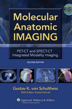
  • Molecular Anatomic Imaging

    20,000.00 9,700.00

    This fully updated Second Edition is perfect for clinicians, as it focuses sharply on clinical PET-CT and SPECT-CT examinations, omitting lengthy physics discussions. The book is now strictly disease oriented and integrates PET-CT and SPECT-CT applications completely. When both techniques are relevant for a disease, they are discussed together; when only one is relevant, it is discussed alone. More than 1,200 illustrations document the use of integrated imaging and provide very useful reference material for interpreting integrated imaging studies.

    A bound-in DVD contains over 80 cases to be viewed in three orthogonal planes and different CT windows organized as reference and self-assessment files. The cases provide excellent training and self-assessment material. Readers can use the DVD to enhance the book’s illustrations or to test their abilities in making diagnoses on their own.

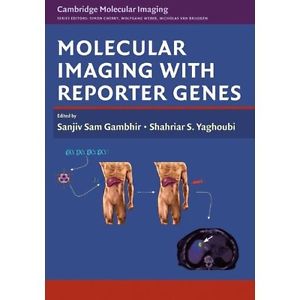
  • Molecular Imaging I

    31,920.00 6,400.00

    The aim of this textbook of molecular imaging is to provide an up to date review of this rapidly growing field and to discuss basic methodological aspects necessary for the interpretation of experimental and clinical results. Emphasis is placed on the interplay of imaging technology and probe development, since the physical properties of the imaging approach need to be closely linked with the biologic application of the probe (i.e. nanoparticles and microbubbles). Various chemical strategies are discussed and related to the biologic applications. Reporter-gene imaging is being addressed not only in experimental protocols, but also first clinical applications are discussed. Finally, strategies of imaging to characterize apoptosis and angiogenesis are described and discussed in the context of possible clinical translation.

  • Multi-slice and Dual-source CT in Cardiac Imaging

    14,995.00 1,990.00

    This book discusses the state-of-the-art developments in multi-slice CT for cardiac imaging as well as those that can be anticipated in the future. It is a comprehensive work covering all aspects of this technology from the technical fundamentals to clinical indications and protocol recommendations. This second edition draws on the most recent clinical experience obtained with 16- and 64-slice CT scanners by world-leading experts. The book also has chapters on area-detector CT and the brand new dual-source CT.

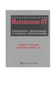
  • Multidetector Computed Tomography: Principles, Techniques, and Clinical Applications

    12,530.00 8,400.00

    Written by the acknowledged experts, this book guides practitioners through the technical aspects of multidetector computed tomography (MDCT) and its site-specific clinical applications in the chest, gastrointestinal organs, genitourinary tract, vascular system, and musculoskeletal system. More than 1,400 illustrations complement the text.

    The contributors explain the advantages of MDCT over single-slice CT in specific clinical situations and offer practical guidance for optimizing scanning protocols. State-of-the-art coverage includes 3-D rendering techniques, the virtual colonoscopy, CT angiography, and screening. Protocols for pediatric patients are included.

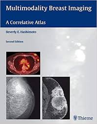
  • Multimodality Breast Imaging: A Correlative Atlas

    15,200.00 8,990.00

    The second edition of this generously illustrated case-based reference provides a systematic visual collection of pathologic entities and a detailed assessment of how to optimize sonographic technique as well as how to approach the integration of mammography, sonography, MRI and PET/CT in breast cancer diagnosis. The book begins with a focus on teaching practical methods to analyze and incorporate mammographic, sonographic, and magnetic resonance findings in the clinical setting. The closing chapters are devoted to illustrating the applications of PET as demonstrated by specific clinical cases.

    Features of the second edition:

    • Emphasis on the importance of high-resolution sonography
    • Three new chapters on the use of MRI and PET in breast imaging
    • Numerous new case studies — including helpful pearls and pitfalls — that focus on common and uncommon examples of metastatic and non-metastatic disease
    • Charts and outlines that provide rapid reference for the clinical workup of a lesion
    • More than 800 images that help identify both mammographic and sonographic abnormalities

    This thorough reference is ideal for radiologists, mammographers, oncologists, gynecologists and all clinicians looking to broaden their visual sonographic experience. Its user-friendly format makes it a handy text for radiology residents in breast rotations.

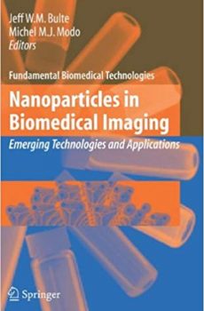
  • Nanoparticles in Biomedical Imaging

    Fundamental Biomedical Technologies features titles in multidisciplinary, technology-driven areas, providing the foundations for breakthrough advances in medicine and biology. The term technology refers, in a vigorously unrestrictive sense, to a broad array of engineering disciplines, the sciences of computation and informatics, mathematical models exploiting and advancing methods of mathematical physics, and the development of novel, experimental discovery devices. Titles in this series are designed and selected to provide high-level visionary input for specialists, while presenting overviews of emerging fields for those in related areas. Volumes in this series aim to provide technologists with the material to gain competent entry into biomedical research and biomedical researchers to understand and embrace novel technological foundations and tools.

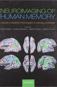
  • Neuroimaging in Human Memory

    10,900.00 3,995.00

    In the past 20 years, neuroimaging has provided us with a wealth of data regarding human memory. However, to what extent can neuroimaging constrain, support or falsify psychological theories of memory? To what degree is research on the biological bases of memory actually guided by psychological theory?

    In looking at the close interaction between neuroimaging research and psychological theories of human memory, this book presents a state-of-the-art exploration of imaging research on human memory, along with accounts of the significance of these findings with regard to fundamental psychological questions. The book starts with a summary of some of the conceptual problems we face in understanding neuroimaging data. It then looks at the four areas of human memory research that have been most intensively studied with modern brain imaging tools – Learning and consolidation, Working memory control processes and storage, Long-term memory representations, and Retrieval control processes. Throughout, the book shows how brain imaging methods, such as functional magnetic resonance imaging (fMRI) and electroencephalography (EEG), can help us increase our knowledge of how human memory is organized, how memory representations are stored, consolidated and retrieved, and how access to memory contents is controlled. With all chapters written by leading researchers in the field, the book will be essential for all those interested in the psychology and neuroscience of memory.

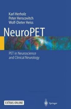
  • NeuroPET

    7,900.00 4,590.00

    This is the only comprehensive and up-to-date book on neurological PET. Its goal is to facilitate cooperation between PET experts, neuroscientists and clinicians. We review PET in neuroscience with particular emphasis on findings that indicate its potential for improving diagnosis and treatment in neurology and psychiatry. We want to improve the transfer of the huge scientific developments in brain PET into clinical care to produce tangible human benefit. We therefore focus on practical and potentially clinically relevant issues and identify solid ground as well as open questions. In addition, the book includes major chapters on the methodological background, including tracer physiology and kinetic modeling, and a comprehensive literature review. The book contains a CD-ROM with video clips and interactive navigation in 3D data sets that illustrate the information contained in PET images in major neurological diseases and its potential to improve diagnosis and neurosurgical planning in brain tumors.

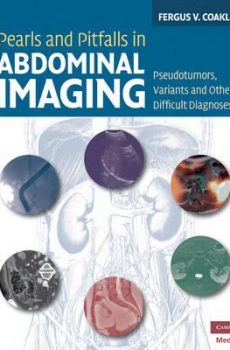
  • Pearls and pitfalls in abdominal imaging

    10,400.00 4,795.00

    Research consistently suggests that 1.0 to 2.6% of radiology reports contain serious errors, many of which are avoidable, and it is clear that all radiologists can struggle with the basic questions as to whether a study is normal or abnormal. Pearls and Pitfalls in Abdominal Imaging presents over 100 conditions in the abdomen and pelvis which can commonly cause confusion and mismanagement in daily radiological practice, providing a focused textbook that can be readily used to avoid wrong diagnoses and prevent incorrect management or even malpractice litigation. It includes 700 figures and covers all the major modalities including CT, PET/CT and MRI. The pearls and pitfalls include artifacts, anatomic variants, mimics, and a miscellany of diagnoses that are under-recognized or only recently described. Conditions are presented in anatomic order from diaphragm to the symphysis pubis, with grouping by location and organ system.

  • Percutaneous Vertebroplasty and Kyphoplasty

    14,990.00 4,590.00

    This new edition of the well-regarded original has been thoroughly revised by Drs. John M. Mathis, Hervé Deramond, and Stephen M. Belkoff to reflect recent advances in percutaneous vertebroplasty and kyphoplasty. Obsolete sections have been judiciously replaced with cutting-edge material, such as an in-depth look at the latest bone cements and devices. Chapters outline spine anatomy, medical management, and patient selection. The addition of practical and challenging case studies furthers the focus of the previous edition by bridging the gap between theory and practice for spine interventionalists, radiologists, neuroradiologists, orthopedic surgeons, and neurosurgeons. The text is enhanced by a wealth of illustrations.

    Featured in the Second Edition:

    * New data on alternate routes for therapy, sacroplasty, and treating tumors

    * New treatment techniques

    * Updated examination of biomechanics

    * New material on complications

    * New figures and color images

    * Inclusion of vertebroplasty and kyphoplasty cases

    * Expanded presentation of ACR and SIR care standards

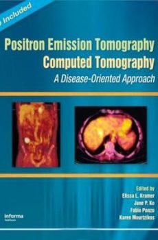
  • Positron Emission Tomography-Computed Tomography

    29,330.00 15,190.00

    Positron Emission Tomography Computed Tomography: A Disease-Oriented Approach offers Radiologists and Nuclear Medicine specialists a thorough understanding of the clinical application of PET-CT―a groundbreaking modality that provides a powerful fusion of imaging anatomy and metabolic function. Written with a disease-oriented approach, PET-CT examines understanding, using, and interpreting PET-CT imaging in clinical practice. Co-authored by experts in both PET and CT imaging, this text serves as an integrated review of the practical aspects of this new imaging modality while providing comprehensive and evidence-based coverage. This volume covers all clinical entities for which PET-CT can be utilized in today’s modern practice.

    Using an integrated disease-oriented approach, PET-CT reviews:

    • the diagnostic settings in which PET-CT will prove most valuable
    • literature-based evidence for utility, applications, and limitations to each disease
    • integrated discussion of the CT findings that will bear on the PET interpretation and vice versa
    • “next steps” in the clinical evaluation of a patient (i.e., additional imaging studies indicated)

    Positron Emission Tomography Computed Tomography also includes a CD packed with every image from the book. With over 340 high resolution photos this makes a perfect addition to both in-depth study and PowerPoint slide presentations.

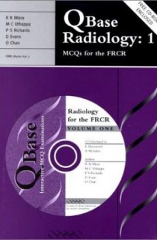
  • Qbase Radiology 1

    3,520.00 2,320.00

    QBase Radiology 1 and 2 together represent a completely comprehensive resource for all postgraduate and undergraduate trainees in radiology. In combination, the books include 900 multiple choice questions (480 on Volume 1 and 420 on Volume 2) and the free CD-ROMs that accompany both books also contain all 900 MCQs and over 100 high quality radiological images. The powerful QBase examination analysis software allows the reader to attempt the same exams as printed in the book, but on screen, realistically replicating the examination situation. The user can also ‘custom-build’ their own exams based on their own choice of subjects and number of questions, sit an exam that is automatically set by the program using all of the questions on the CD, or they can sit a ‘shuffled’ version of an exam, whereby the question parts (A-E) are reorganised automatically to avoid pattern recognition. The software also allows the user to set their own ‘confidence level’ when taking an exam and it will automatically mark, analyse and store completed exams for future review and re-sitting, allowing the package to be used as a fully interactive self-learning tool.

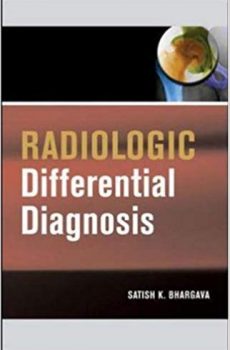
  • Radiologic Differential Diagnosis

    4,620.00 2,990.00

    This is the perfect quick-reference for the busy radiologist and resident. The most up-to-date resource in the field of radiography, this bulleted handbook provides differentiating features for hundreds of diseases, conditions, and injuries. Readers will find 300 illustrations and radiographs, a streamlined find-it-now format, and exceptional value for the price.

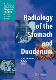
  • Radiology of the Stomach and Duodenum

    16,990.00 6,790.00

    A number of imaging techniques, many of them complementary, are used in the investigation and treatment of disorders of the stomach and duodenum. Optimal patient treatment requires a thorough knowledge of the application of these techniques, as well as a sound understanding of pathology of the stomach and its presenting symptomatology. This well-illustrated book covers the various investigative methods in detail, discussing their advantages and disadvantages and explaining their role in specific settings. It will be of great value to both trainee and experienced radiologists, and should assist in promoting effective and judicious patient management.

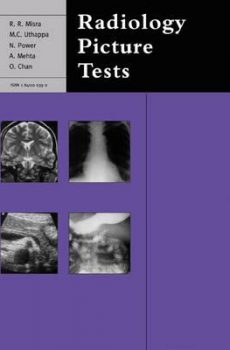
  • Radiology Picture Tests

    3,900.00 2,700.00

    A comprehensive, systemically organised ‘film viewing’ book designed specifically for FRCR candidates but equally useful to all trainee and junior radiologists as an ‘aide memoire’ and to medical undergraduates and MRCP candidates as a useful means of self-testing their level of knowledge. It is fully illustrated with high-quality, clearly labeled black and white illustrations throughout and the text closely mirrors the way that questions are asked in the FRCR film viewing examination, so as to provide as real an examination experience as possible. Each section of the book contains 20 representative questions, and model answers, providing 200 cases altogether.

Contact Info

Calicut Books
Opp.Govt Mental Health Centre,
P.O Kuthiravattom,
Calicut - 673016

0495 - 2740321 +91 9747580707

© Copyright Calicut-Books 2017 All Rights Reserved